
Cytology is the study of cells, their structure and function. It is an important area of biology, providing vital insights into the workings of life at the most basic level. Through cytological analysis, scientists can learn about the process of cell division, development and death, as well as how cells interact with each other in order to form tissues and organs. Cytology is also used to identify abnormal cells in cancer diagnosis and treatment, making it a key tool for medical research.
It involves examining a sample, often from a biological fluid or tissue, using specialized equipment such as a microscope or cytology centrifuge. Using these tools, scientists can identify abnormalities in cells that could indicate illness or disease.
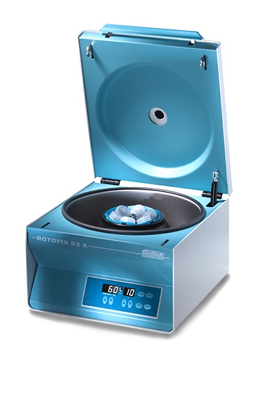
What is a Cytology Centrifuge?
Centrifuges are a key tool in cytology laboratories. They enable scientists to transfer cells onto microscope slides, allowing for the identification and analysis of cells. Cytology centrifuges help speed up processes such as cell counting and cell sorting.
Most modern cytology centrifuges consist of an outer casing containing a rotor to spin the samples, a motor to power the rotor, and various safety features. The rotor is usually made from lightweight aluminum or plastic and may have either an open or closed design.
The motor is designed to provide a certain amount of acceleration and deceleration in order for the samples inside to be spun at a set rate. Safety features include a lid interlock to prevent the rotor from spinning when the lid is open, as well as various sensors and alarms that alert users to any potential issues.
How a Cytology Centrifuge Works?
The samples to be spun in a cytology centrifuge are usually placed into small funnels. These funnels contain either an aqueous solution or a suspension of the sample to be observed. Once the funnels are loaded into the centrifuge, the rotor is turned on and it spins at a certain speed, depending on the type of sample being processed.
As the rotor spins, it creates an outward force on the funnels that causes cells to be transferred onto a microscope slide.
Once the centrifugation process is complete, the samples can then be stained and analyzed using microscopy By spinning cells inside a cytology centrifuge, scientists can quickly and accurately prepare cells for further study.
In short, cytology centrifuges are an essential tool in the laboratory for preparing cells for analysis under a microscope. They allow researchers to easily identify and analyze cells so that they can better understand a variety of biological processes.
With their ability to accelerate and decelerate quickly, cytology centrifuges are an efficient tool in the laboratory.